Keratoconus treatment in Delhi at Jain Eye Hospital & Laser Centre, Shalimar Bagh, depends on how far the cornea has changed. Options run from glasses and special lenses to corneal cross-linking (C3R), rings, laser with cross-linking, ICL and transplant. Fees depend on the procedure and examination. The C3R and INTACS fees are marked Confirm pending written confirmation from the hospital. Dr Rajat Jain reviews this page.
Medical reviewer: Confirm. Reviewer registration and source check: Confirm. Last reviewed: Confirm.
Book a keratoconus assessment or call 9643 900 900.
What is keratoconus?
The cornea is the clear front window of the eye. In keratoconus its collagen framework weakens, and the cornea slowly thins and bulges forward into a cone. A cone-shaped cornea bends light unevenly, so vision becomes blurred, doubled or streaked, and glasses stop correcting it well.
It usually starts in the teens or twenties and often affects both eyes, though one eye may be worse. Most people notice it between the ages of about 15 and 30. It can stabilise on its own in the thirties, but nobody can tell in advance whether a particular eye will do so, and that is the reason for scans at intervals rather than a single visit.
Keratoconus is not cancer, it is not an infection, and it is not caused by wearing glasses or by screens. Genes, eye rubbing and allergic eye disease are the most commonly named contributors. It is more often found when it runs in the family.
What are the early signs of keratoconus?
Early keratoconus can be quiet. These are the signs that deserve a corneal scan:
- Glasses power, especially cylinder (astigmatism), that keeps changing every few months
- Ghost images, or one image seen as two or three, especially with one eye covered
- Streaks, starbursts and glare at night
- Vision that is never quite sharp even with new glasses
- A strong urge to rub the eyes, often with itching
- A sudden misty patch with pain, which can follow a break in the inner layer (hydrops)
Your optician may notice a change in the reflex during testing, but only a corneal scan can show the shape. If someone in your family has keratoconus, screening a teenager early is sensible even when vision seems normal. Always rule it out before laser vision correction; our LASIK surgery page explains why.
How is keratoconus diagnosed?
Diagnosis rests on mapping the front and back surface of the cornea and measuring its thickness. A corneal topography or tomography scan takes a few seconds per eye and does not touch the eye. Confirm The scan shows where the cornea is steepest, how thin it is at the thinnest point, and how the back surface behaves.
A typical assessment also includes:
- A glasses test (refraction), and a slit-lamp examination of the cornea
- A dilated check of the retina and optic nerve
- Thickness measurement (pachymetry)
- A tear film check, because dry eye and allergy affect both the scan and the plan
- A repeat scan after a few months if the first one is borderline, to see whether the shape is changing
Progression means a measurable steepening or thinning between scans, or a rising cylinder in glasses. That finding, rather than the label alone, usually decides when cross-linking is advised. The visit takes about an hour.
Which treatment suits which stage, and what does each cost per eye?
Keratoconus is treated in steps. The aim in early disease is to stop progression and give the clearest vision with the least intervention. The aim in later disease is to restore useful vision.
| Option | What it does | What it does not do | Usual stage | Fee per eye |
|---|---|---|---|---|
| Glasses | Corrects mild blur and cylinder | Does not stop progression | Early, mild | Not applicable (optical shop) |
| Soft or hybrid lenses; rigid gas-permeable (RGP) and scleral lenses | Make a smooth new surface in front of the cone and give sharper vision | Do not stop progression; need fitting, care and follow-up | Mild to advanced | Confirm |
| Corneal cross-linking (CXL, C3R) | Riboflavin drops and ultraviolet light strengthen the collagen, aiming to stop the cone from advancing | Does not reverse the cone; vision may stay blurred, and glasses or lenses are often still needed | Early to moderate, when progression is shown | Confirm |
| Intracorneal ring segments (INTACS / ICRS) | Small arcs placed in the cornea flatten and regularise it; may improve lens tolerance and vision | Do not stop progression on their own; cross-linking is often added | Moderate, when lenses are not tolerated | Confirm |
| Topography-guided PRK with cross-linking | A surface laser smooths the irregular shape, and cross-linking holds it | Not for thin or advanced corneas; vision correction is partial | Selected mild to moderate, with adequate thickness | Confirm |
| ICL (Implantable Collamer Lens) | A lens inside the eye corrects high power without touching the cornea | Does not treat the cone itself; only for a stable cone | Stable keratoconus with high power | Confirm |
| Corneal transplant (DALK or PK) | Replaces the diseased cornea with donor tissue | Does not remove the need for glasses or lenses afterwards; takes months to settle | Advanced, scarred or lens-intolerant | Confirm |
These are indicative ranges. The exact fee is confirmed after examination, because the procedure, the lens or drug used and the condition of your eye decide it. Fees last updated [month year, CONFIRM]. Pre-operative tests, medicines and follow-up are billed separately unless stated. Cornea transplant details, including donor tissue, are on our cornea treatment page. All other fees are on treatment costs. Confirm
In practice the plan combines steps. A teenager with changing cylinder may have cross-linking first and glasses afterwards; an adult with a stable cone and poor lens tolerance may need rings; an older person with a scar may need a transplant. The doctor explains which step comes first and why.
Who should not wait?
Cross-linking works better before the cornea has thinned or scarred. These are the situations where waiting costs vision:
- A teenager or young adult with a rising cylinder or a scan showing steepening
- A child with keratoconus, because it tends to progress faster at younger ages
- Strong eye rubbing or allergic eye disease that is not yet controlled
- A second eye that is clearly changing after the first eye needed treatment
- A cornea close to the thickness limit for treatment, which gets thinner as time goes on
If you are 35 or older with a scan that has been unchanged for years, watching and lenses may be enough. The choice is made on repeat scans, not on age alone.
What happens on the day of cross-linking (C3R)?
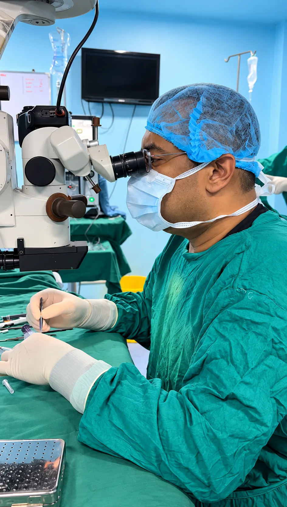
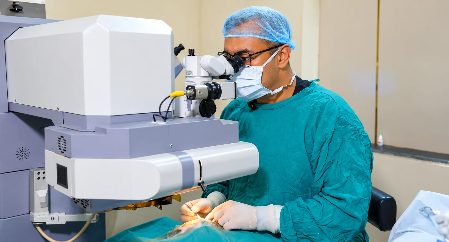
Standard cross-linking is a day-care procedure on one eye at a time, and it takes about an hour in the theatre. Your protocol may differ Confirm.
- Numbing drops are put in and the eyelids are held open with a small holder.
- In the standard method, the thin surface layer (epithelium) is gently removed so that the riboflavin can enter the cornea.
- Riboflavin (vitamin B2) drops are applied every few minutes for around 30 minutes until the cornea is saturated.
- The cornea is exposed to controlled ultraviolet-A light for a set time, with more drops during exposure.
- A bandage contact lens is placed on the eye to protect it while the surface heals, along with antibiotic and other drops.
- You rest briefly and go home with a companion.
Most people feel little during the procedure, though the evening after can be uncomfortable, and the second eye is usually treated weeks or months later if it needs it.
How long is recovery after cross-linking?
| When | What to expect | What to do |
|---|---|---|
| First 24 to 48 hours | Burning, watering, light sensitivity; the worst of the discomfort | Rest, drops as written, dark glasses, painkiller as advised by the doctor |
| Days 3 to 5 | The surface heals; discomfort eases | Review visit; the bandage lens is usually removed around day 4 to 7 |
| Weeks 1 to 4 | Vision may be hazy, with glare | No rubbing, no swimming, no dusty or smoky places; drops continue |
| Months 1 to 3 | Haze fades; vision slowly stabilises | Repeat scans; glasses or lenses are re-measured |
| Months 6 to 12 | Cornea settles; the scan shows whether progression has stopped | Further treatment if needed |
Most people take two to five days off work or college. Delhi dust and smoke during winter and Diwali can irritate a healing surface, so keep goggles on if you ride a two-wheeler. Do not rub your eyes, however itchy they feel, and tell us if allergy flares.
What are the risks of each treatment?
- Cross-linking. Temporary pain and haze are common. Infection, delayed healing of the surface and a lasting scar are uncommon but real. In some eyes the cone still progresses, and a second treatment is occasionally needed.
- Rings. Ring displacement or exposure, glare and halos, and the need for lens fitting afterwards. They do not cure the cone.
- PRK with cross-linking. Haze, a slower and more uncomfortable recovery than cross-linking alone, and partial correction only.
- ICL. Infection inside the eye, cataract and raised pressure are rare risks of any lens placed inside the eye. It needs a stable cornea.
- Transplant. Rejection, infection, high pressure and the need for months of drops and visits. Recovery is slow.
No success percentages are quoted here because they differ by stage, age and protocol; the surgeon discusses what applies to your eye.
Come in the same day if: you have a sudden misty patch of vision with pain in an eye with keratoconus, new redness and pain after cross-linking or a transplant, vision that suddenly falls, or discharge from the eye. Call 9643 900 900. See eye emergency.
Does eye rubbing and allergy cause keratoconus to progress?
Rubbing is one of the well-established triggers for progression, and allergic eye disease is a common reason for rubbing. Each hard rub presses on a weakened cornea. If your eyes itch, treat the allergy rather than the itch: cool compresses, lubricants and the drops your doctor prescribes. Children with spring catarrh need particular attention because of the intense itch. Our eye allergy page explains the treatment, and dry eye treatment helps if a gritty surface is part of the problem.
Rubbing the eyes with a knuckle, sleeping with the face pressed into the pillow and rubbing during screen use are habits worth breaking. Steroid drops used without advice are not a solution.
Does insurance cover keratoconus treatment?
It depends on the policy. Some insurers cover cross-linking or transplant when it is medically necessary and documented, and others exclude it or put a cap on cosmetic or optical items. Rings and ICL are more often questioned. Cashless eligibility and available services: Confirm with the hospital and insurer, and our desk reads the policy with you before treatment. See insurance and payment. Keep the scans, because insurers often ask for proof of progression.
Keratoconus treatment in Shalimar Bagh for patients across Delhi
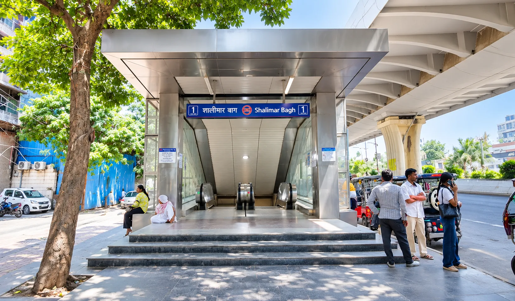
We offer keratoconus treatment in Shalimar Bagh at AG-152, Poorbi Shalimar Bagh, near Richi Rich Banquet, Delhi 110088, close to the Shalimar Bagh Metro station on the Pink Line Confirm. Students and families from Rohini, Pitampura, Model Town, Ashok Vihar, Paschim Vihar and nearby areas often come for the first scan before an exam break, and then for scans at intervals. Plan about an hour for the assessment, and bring your latest glasses, any lens prescription and old scans. Confirm Directions are on our Shalimar Bagh hospital page and travel details by neighbourhood are on the North West Delhi page.
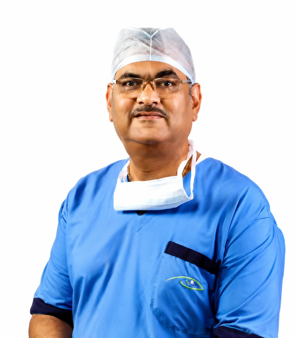
Dr Rajat Jain and your keratoconus assessment
Doctor-specific title, qualifications, registration, clinical assignments, procedures and OPD availability: Confirm. See his doctor profile or read more on Dr Rajat Jain's own site. If you are looking for keratoconus treatment in Delhi, book a keratoconus assessment. Parents should bring a child for scanning if a sibling or parent has it.
FAQs
Can keratoconus be cured?
Keratoconus cannot be reversed, but corneal cross-linking can slow or stop its progression in many eyes, especially when done early. Glasses, rigid or scleral lenses, rings and, in advanced cases, a corneal transplant help restore useful vision. Regular scans show whether the cone is changing and when treatment is needed.
What is C3R or corneal cross-linking?
C3R is a short day-care procedure in which riboflavin drops and controlled ultraviolet light strengthen the corneal collagen so that the cone is less likely to advance. It aims to stabilise the cornea rather than to improve vision. The current fee is Confirm.
How much does keratoconus treatment cost in Delhi?
At Jain Eye Hospital, per eye: cross-linking Confirm; INTACS Confirm; topography-guided PRK with cross-linking 85,000 to 1,00,000; ICL 70,000 to 1,20,000+; DALK or PK transplant 80,000 to 1,50,000+. These are indicative ranges, and the exact fee is confirmed after examination.
Is cross-linking painful?
The eye is numbed, so the procedure itself is mostly pressure and a feeling of being watched under a light. The evening and next day can be uncomfortable, with burning, watering and light sensitivity, as the surface heals. The pain usually eases within two to three days with drops and rest, and persisting pain should be reported.
Will I still need glasses after cross-linking?
Often yes. Cross-linking aims to stop the cone from advancing, not to correct vision. Many people still need glasses or lenses afterwards, and vision may become a little clearer over several months. Some need rings or laser later. The doctor explains what to expect for your stage at the time of planning.
At what age does keratoconus stop progressing?
It tends to progress most in the teens and twenties and slows in the thirties or forties, but it varies. There is no age at which you can safely stop watching it. Repeat scans every few months at first, and then yearly, show whether your cornea is changing and whether treatment is needed.
Is keratoconus hereditary?
It sometimes runs in families, but most cases appear without a clear family history. Having a parent or sibling with keratoconus raises the chance, so brothers, sisters and children are advised to have a corneal scan from the teenage years. Eye rubbing and allergy add to the risk and can be controlled.
Can I have LASIK if I have keratoconus?
No. LASIK thins the cornea and can make keratoconus worse, so it is not advised in keratoconus or suspected keratoconus. Pre-LASIK scanning is designed to find it. For some stable cases, other options such as an ICL, or topography-guided PRK with cross-linking, may be considered after assessment.
When is a transplant needed for keratoconus?
A transplant is considered when the cornea is too thin, scarred or steep for lenses to give useful vision, or after hydrops leaves a dense scar. Most people with keratoconus never need one. A deep anterior lamellar transplant (DALK) keeps the patient's own inner layer, and penetrating keratoplasty (PK) replaces the full thickness.
Can children have cross-linking?
Yes, when scans show progression. Keratoconus often moves faster in children and teenagers, so treatment is usually advised sooner than in adults. Children need an examination under comfortable conditions, strict instruction not to rub, and treatment of any allergy. The doctor discusses timing with the parents.