

Eye hospital technology in Delhi ranges from simple pressure checks to lasers and scans. This page names the main equipment at Jain Eye Hospital & Laser Centre, Shalimar Bagh, for laser vision correction, cataract surgery, retina, glaucoma and cornea, and says what each is for, who needs it and how to prepare. Every model is Confirm.
Hospital reviewer: Confirm. Last reviewed: Confirm.
What equipment does Jain Eye Hospital use, and why does it matter?
A machine does not make a diagnosis. Your doctor does, using what the machine measures. We list equipment for two practical reasons: so you know which tests you may be asked to have, and so that you can compare what a surgeon proposes with what the hospital can actually do on site. Make and model names are added only after the hospital confirms them.
Which test or machine is used for what?
| Test or machine | Used for | Department | How to prepare |
|---|---|---|---|
| Excimer laser Confirm | Reshaping the cornea in LASIK and PRK | Refractive | Stop contact lenses as instructed; no eye make-up or perfume on the day |
| Corneal topography or tomography Confirm | Mapping corneal shape; screening for keratoconus | Refractive, cornea | Stop contact lenses for the number of days the hospital advises Confirm |
| Pachymetry Confirm | Measuring corneal thickness | Refractive, cornea | None |
| Phaco machine Confirm | Breaking up and removing a cataract through a small cut | Cataract | Follow the fasting and medicine instructions you are given |
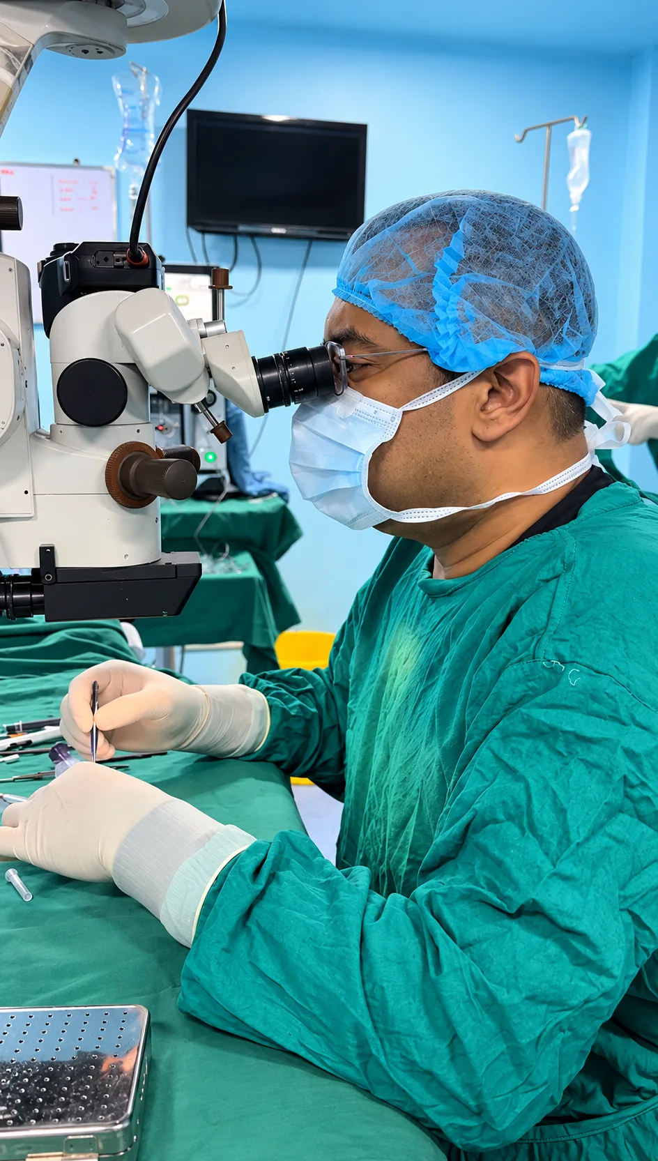
| Operating microscope Confirm | Magnified view during surgery | Cataract, cornea, retina | Not applicable |
| Biometry Confirm | Measuring eye length to choose lens power | Cataract | Stop contact lenses as advised; keep still and look at the light |
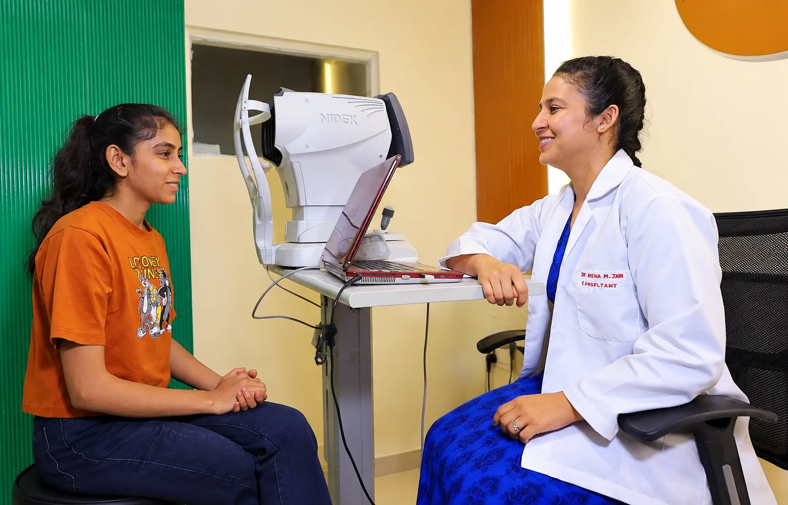
| Tonometer Confirm | Eye pressure | Glaucoma, general | Do not rub the eye; numbing drops may be used |
| Visual field analyser Confirm | Mapping side vision | Glaucoma | Wear your reading glasses if advised; allow 20 to 30 minutes |
| OCT scanner Confirm | Cross-section scan of retina, macula and optic nerve | Retina, glaucoma | Often done without dilation; bring earlier scans for comparison |
| Fundus camera Confirm | Photographs of the retina | Retina, diabetic eye check | Dilating drops may be used; bring a companion |
| Retinal laser Confirm | Sealing tears and treating diabetic retinopathy | Retina | Eat normally unless told otherwise; bring a companion |
Preparation steps are general. The hospital confirms the instructions for your visit when you book.
What does the laser used for LASIK and PRK do?
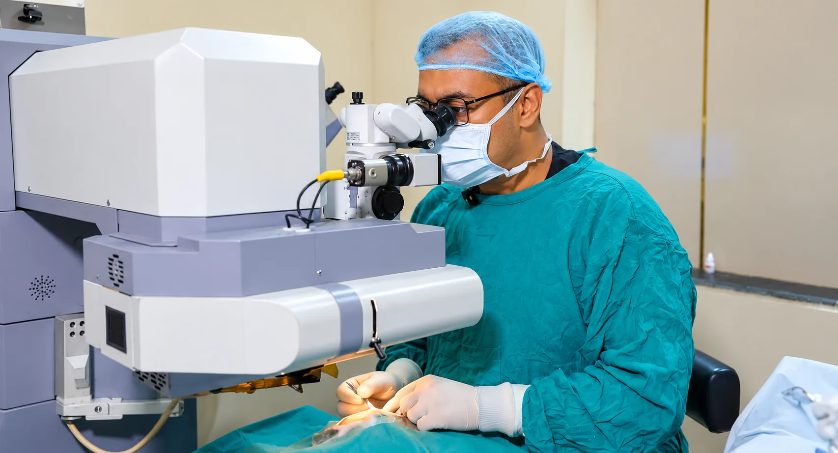
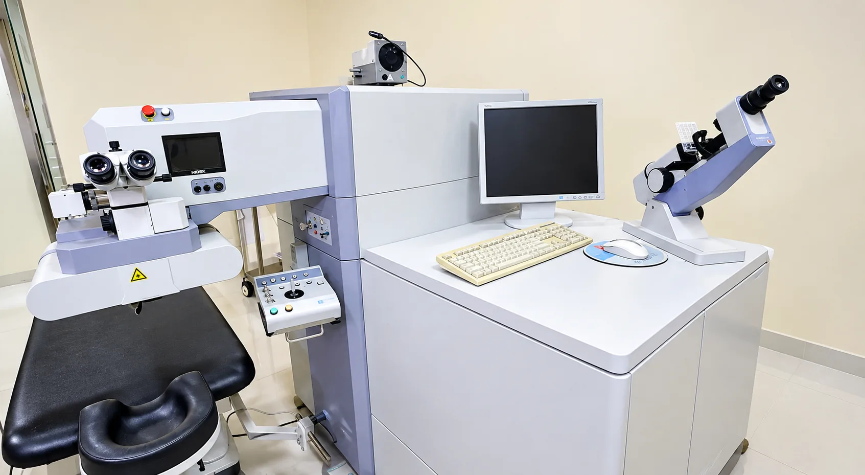
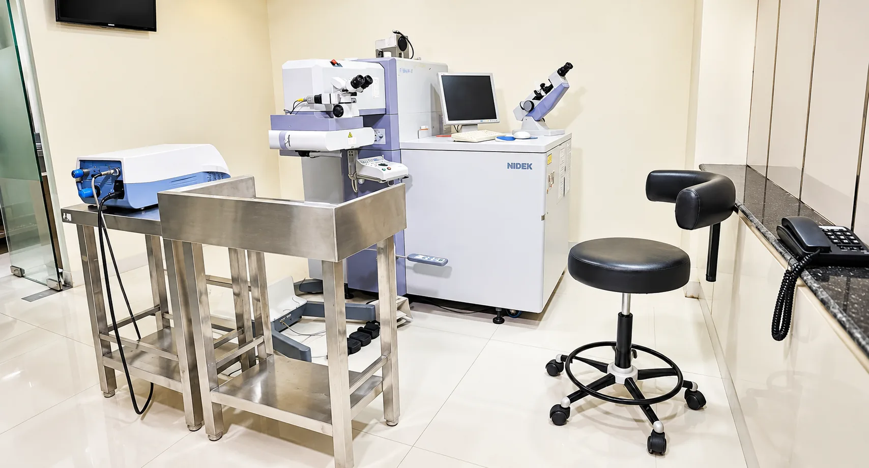
An excimer laser removes a very thin layer of tissue from the cornea to change its focusing power. It is used for short-sightedness, long-sightedness and astigmatism within limits that the corneal tests decide Confirm. Numbing drops are used, you look at a light, and most people feel pressure rather than sharp pain; the laser itself usually runs for a short time per eye. The device matters less than your corneal thickness, shape and stable power, which the tests below check. Read the LASIK surgery page before you book.
What is used in cataract surgery?
Cataract surgery uses ultrasound (phacoemulsification) to break up the clouded lens through a small cut, and the operating microscope to see it. Before surgery, biometry measures the length of the eye and the curve of the cornea, and a formula turns these into the power of the lens to be implanted. A wrong measurement means a wrong lens power, so the test is repeated if the readings look doubtful. Whether a femtosecond laser is part of the service is Confirm. The techniques are compared on our cataract techniques page.
How do retina scans and laser work?
Optical coherence tomography (OCT) shines harmless light and builds a cross-section of the retina, like an ultrasound that uses light. It shows swelling at the macula, fluid, holes and thinning. It takes a minute or two per eye, involves no touch and is repeated at follow-ups to see change. The fundus camera photographs the retina. A retinal laser applies small burns to seal a tear or treat diabetic retinopathy; the patient sits at a microscope with numbing drops and sees bright flashes. See retina treatment and the eye checkup page.
Which glaucoma tests are done?
Glaucoma damages the optic nerve, often with no early symptoms. Three measurements work together: eye pressure (tonometry), the shape and thickness of the nerve (OCT or examination) and the visual field, which maps what you see at the sides. Gonioscopy, a contact-lens look at the drainage angle, is done in clinic Confirm. One high pressure reading does not mean glaucoma, and a normal reading does not rule it out, which is why several tests are used. See glaucoma tests.
How is the cornea imaged?
Topography or tomography maps the front surface (and for some devices the back surface) of the cornea. It picks up irregular shape, such as keratoconus, before you notice it, and is required before laser vision correction. Pachymetry measures thickness. Contact lenses change the shape of the cornea, which is why you stop them before this test. See cornea treatment.
How is the operation theatre kept safe?
Instruments are sterilised by Confirm; single-use items such as blades and cannulas are not reused. The theatre is cleaned between cases Confirm. Ask the front desk to describe what happens to your instruments on the day; you are entitled to the answer. Day-care recovery arrangements: Confirm.
What do we not claim?
We do not say our equipment is the newest, the most precise or the only one of its kind, and we do not compare it with other hospitals. We do not promise an outcome because of a machine. The devices above are tools; suitability for surgery, and the technique chosen, depend on your examination. If a machine you read about is not listed here, it is not part of the service described here Confirm. If you want a test the doctor has not advised, ask why before you pay for it.
Not sure which tests you need? Book an eye checkup or call 9643 900 900. Fees are in treatment costs.
Questions to ask about a test or device
Technology is useful when it answers a clinical question. It does not replace a conversation about your symptoms, examination and choices. Ask the team what a proposed test is called, what part of the eye it examines, why it is being considered for your visit and whether it is needed today or can be discussed later. If you are asking about a particular device or model, confirm its availability before planning your visit.
If a scan or measurement is recommended, ask what you will need to do, how long the appointment may take and whether any preparation is required. Tell the team about previous eye procedures, current drops, contact lenses and relevant earlier reports. Do not stop a medicine, change a drop schedule or change lens wear unless the clinician gives you that instruction. If you have an earlier scan, ask whether it would help to bring it so the doctor can decide whether comparison is useful.
Ask who will explain the result and how it relates to the treatment options being discussed. A device image or measurement is one part of an assessment; ask what it can and cannot show, whether another examination is needed and what would change the recommended plan. If two options are offered, request a plain-language explanation of the expected benefits, limitations, risks and follow-up for each. The doctor should relate that discussion to your own examination rather than promise a result from a device alone.
Before agreeing to a test or procedure, ask for the fee and what it includes. The treatment costs page lists published ranges where supplied, while your written estimate after examination is the figure for your proposed care. Ask whether medicines, repeat measurements, follow-up visits or other items are separate. If an insurer or panel is involved, ask the desk what documents it needs and confirm coverage with the insurer as well.
You can keep a copy of reports and ask how to share them securely with the hospital. Do not send detailed medical records through a general website message. For a routine eye examination, read the eye check-up guide; to request a visit, use the appointment form. If an equipment model or in-house service is not confirmed on this site, call the team rather than assuming that it is available.
FAQs
Which laser is used for LASIK at Jain Eye Hospital?
LASIK and PRK are done with an excimer laser Confirm. It reshapes the cornea to correct minus, plus and cylindrical power within set limits. Your pre-operative tests, including corneal mapping and thickness, decide which procedure suits you, and whether you are suitable at all.
Is OCT available at the hospital?
Confirm. If yes: OCT scans for the retina, macula and optic nerve are done in the hospital, usually on the same visit as the examination. It is a quick scan with no contact with the eye. The doctor advises it only when it will help the diagnosis or follow-up.
Is femtosecond laser cataract surgery available?
Confirm. If not: the hospital does phaco surgery, in which the clouded lens is broken up with ultrasound through a small cut. This technique suits most cataracts. Your doctor explains if your eye needs a different method, and the choice of technique and lens is written into your estimate.
How are instruments sterilised?
Instruments are sterilised by Confirm before every surgery, and single-use items such as blades and cannulas are not reused. The theatre follows infection-control routines including cleaning between cases. The hospital will state the actual protocol once confirmed.