








The cornea: the clear front surface of the eye
The cornea is the transparent front surface of the eye. It helps focus incoming light and protects the structures inside the eye. Its smoothness and clarity matter: swelling, scarring, infection, injury, dryness or an irregular shape can make vision blurred and the eye uncomfortable. Corneal care covers several different conditions, so the first step is to find the cause rather than treating every red or painful eye in the same way.
Keratoconus is a condition in which the cornea becomes thinner and changes shape, which can produce irregular astigmatism and blurred vision. Infection, abrasion, chemical injury, inherited disorders and contact-lens-related problems are separate possibilities. Some problems are minor, while others threaten sight and need urgent treatment. Avoid home remedies, old drops or another person’s contact lenses. An examination allows a clinician to check the surface, measure shape where appropriate, and decide what care is safe.
Symptoms that may involve the cornea
Corneal problems can cause blurred or fluctuating vision, a gritty or foreign-body feeling, watering, light sensitivity, redness or pain. The pattern varies: a small surface scratch can be painful, while a gradual shape change may mainly blur vision. Contact-lens wearers should take new pain, redness, light sensitivity or blur seriously, remove the lens and contact an eye-care professional promptly. Do not put the lens back in until a clinician has advised that it is safe.
Chemical splashes and penetrating injuries are emergencies. For a chemical exposure, begin rinsing the eye immediately with clean, lukewarm running water and seek emergency care; do not delay rinsing while searching for special drops. Do not rub an injured eye or try to remove an embedded object. A painful red eye with declining vision, sudden clouding or discharge should also be assessed urgently. Symptoms are clues, not a way to distinguish infection from allergy or another eye condition at home.
Common corneal problems are not all alike
Keratoconus changes corneal shape and may cause progressive irregular vision. Corneal infection or an ulcer can damage tissue and may need urgent treatment, especially for contact-lens users. Dry-eye disease affects the tear film and surface, causing irritation or fluctuating blur; it can coexist with a separate corneal condition. Scratches and foreign bodies affect the surface after injury. Scars and inherited corneal disorders may alter transparency in different ways. Naming the condition matters because treatment for one problem may worsen another.
Tell the clinician about contact-lens wear and cleaning habits, eye rubbing, allergies, recent trauma, previous infections, eye drops and family history of corneal disease. Eye rubbing may be associated with corneal problems and can aggravate irritation; ask for help managing itch rather than rubbing. Do not self-diagnose keratoconus from a glasses prescription or a family member’s experience. A specialist can say whether a corneal-shape examination or further assessment is indicated.
Examination and corneal-shape measurements
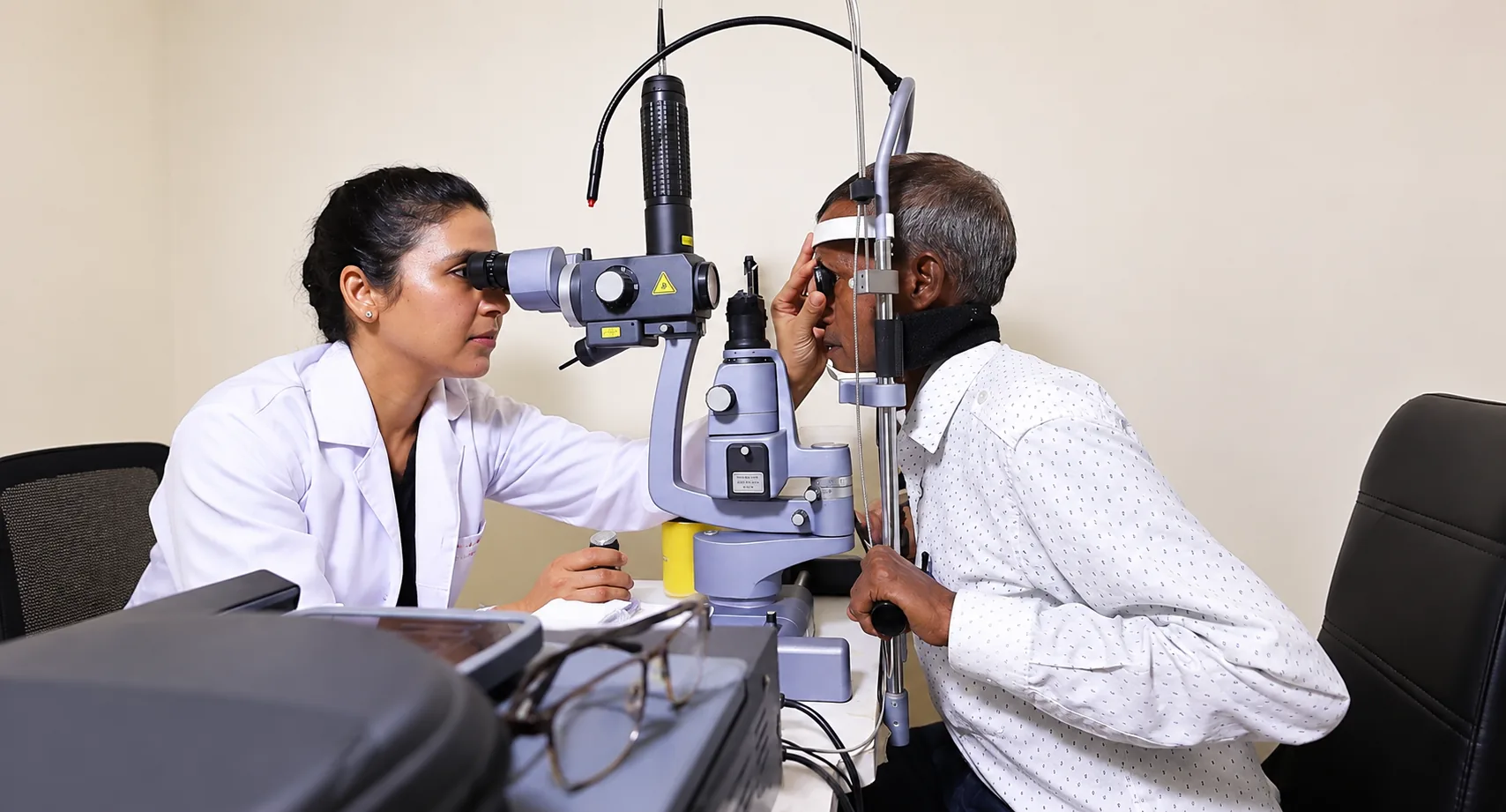
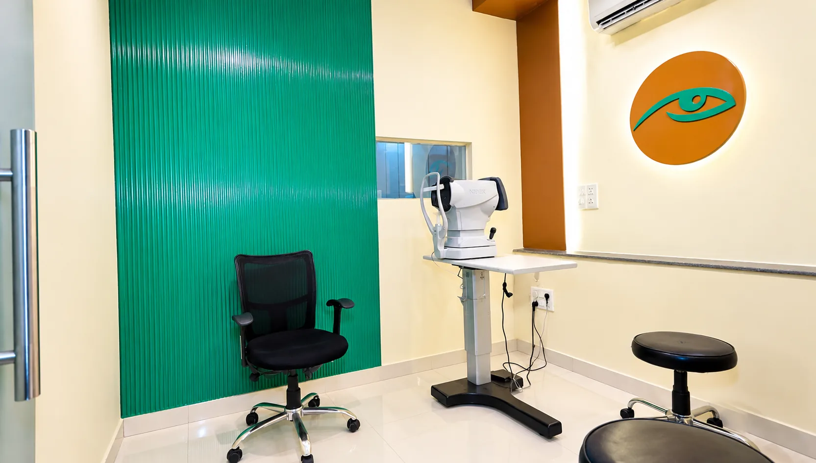
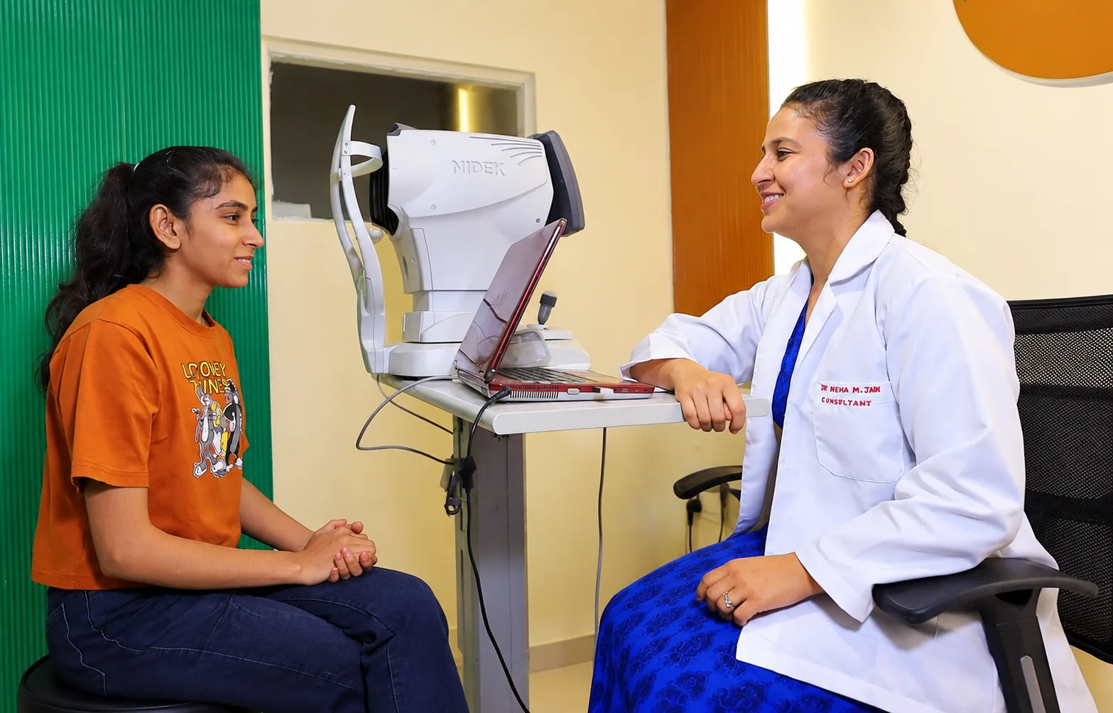
A corneal assessment may include visual-acuity and refraction checks, examination with a slit lamp, staining of the surface, and measurements of corneal curvature or thickness. If keratoconus or another shape disorder is suspected, corneal topography or tomography may be considered. These maps show shape information that a routine glasses test cannot provide. The clinician chooses examinations based on the history and findings; not every patient needs every test.
Bring glasses, contact lenses or their packaging, cleaning-solution details, previous prescriptions and any past corneal maps. Tell the team when lenses were last worn because some may change the corneal surface temporarily; ask whether to pause wear before a planned measurement. If there is an active painful or red episode, mention when it started and what drops have been used. Ask what each test checks, whether both eyes need measurement, and what finding would change the plan.
Treatment is matched to the corneal problem
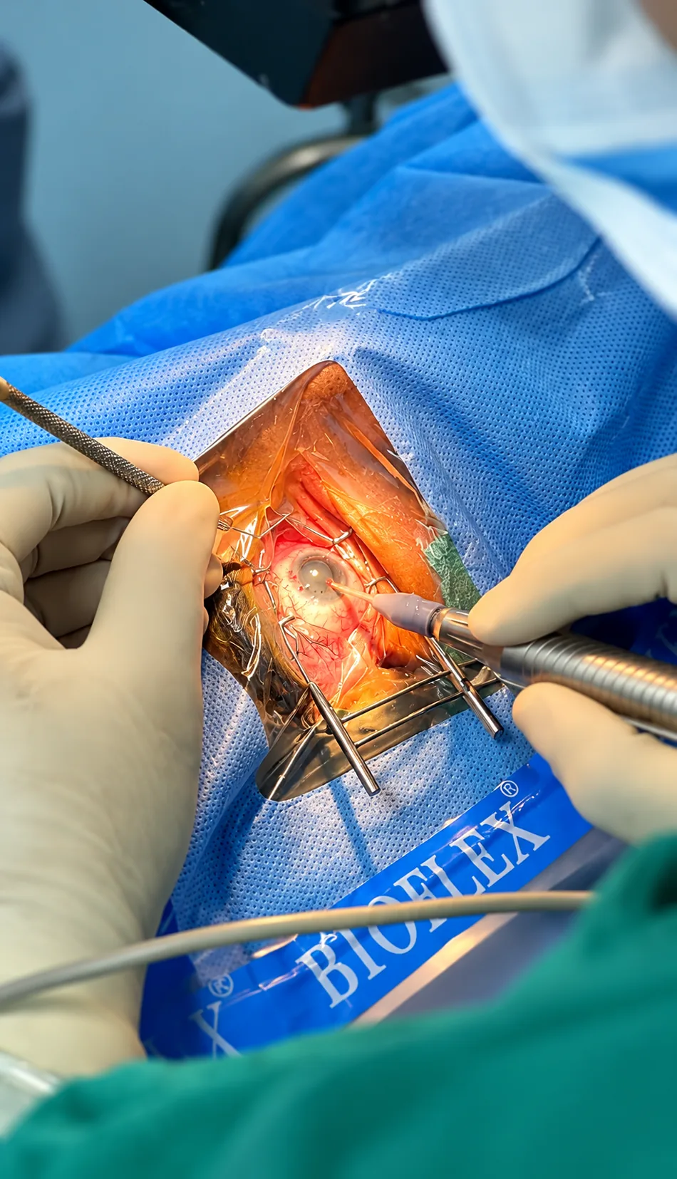
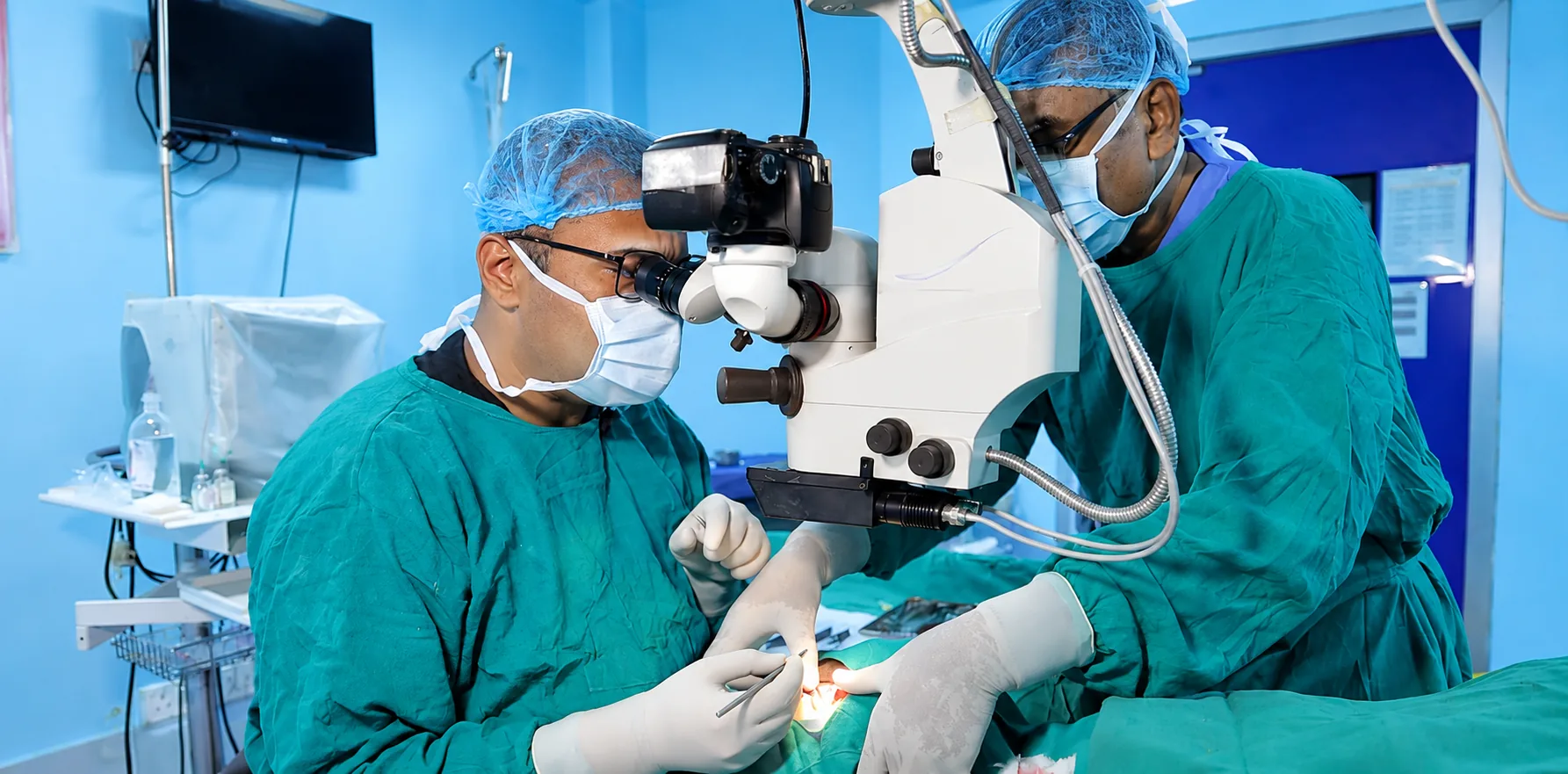
Treatment ranges from lubricating or prescription drops to specialty contact lenses, managing an infection, protecting an injured surface, or considering a procedure for a progressive shape disorder. Keratoconus care may involve spectacles or contact lenses for vision; corneal cross-linking may be discussed to slow progression in suitable cases. Cross-linking is not the same as a vision-correction operation and does not guarantee that glasses or lenses will no longer be needed. A corneal transplant is considered only in selected cases when other care is not adequate.
For infection, the medicine depends on the likely cause; steroid drops can be harmful if used without checking for infection. Do not use leftover antibiotic or steroid drops, and do not share medication. Ask what diagnosis is being treated, how quickly improvement should occur, what risks or side effects matter, and when to return. Contact-lens wear may need to stop until the surface is safe. The clinician should give an individual plan and verify which tests and treatments are actually available at the hospital.
Why corneal maps and surface checks matter
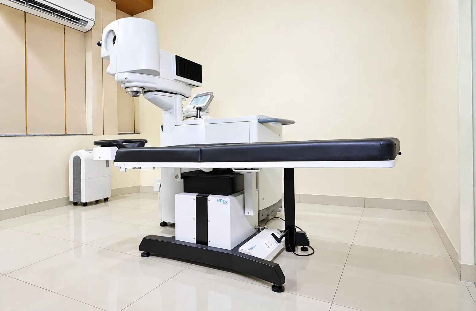
Corneal mapping can help assess irregular shape, compare measurements over time and support planning when keratoconus is suspected. A thickness measurement may be relevant to the safety of some procedures, while slit-lamp examination helps assess transparency and the surface. These measurements are not interchangeable; one normal result does not necessarily answer every question. A clinician interprets them with visual function, symptoms and previous records.
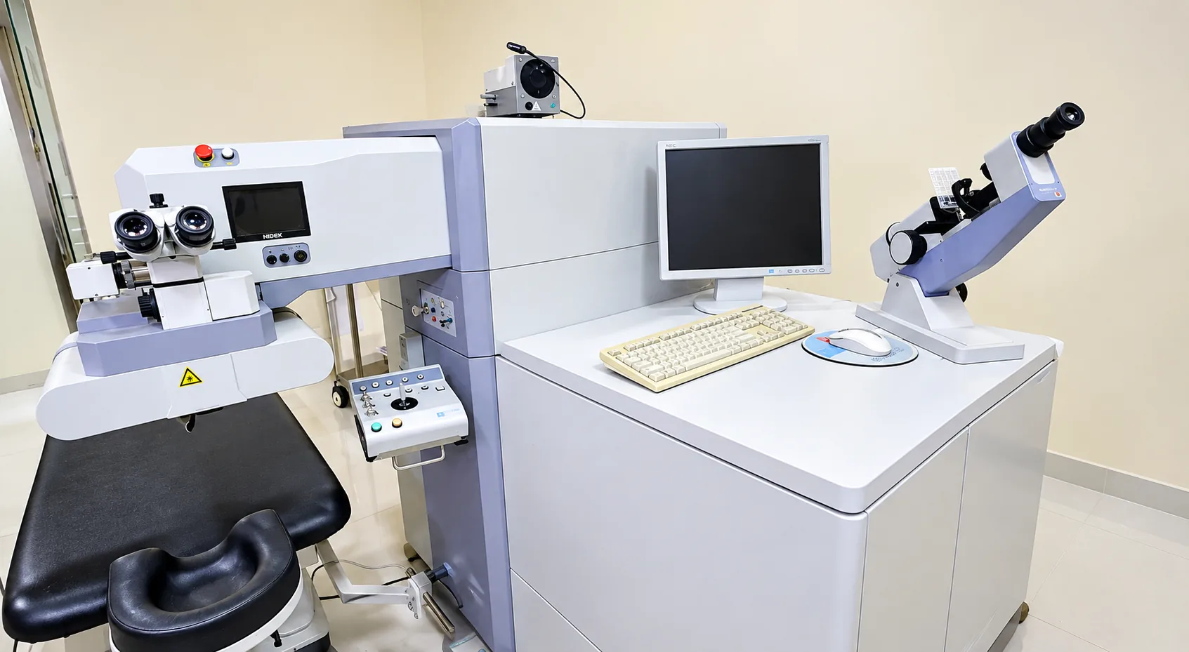
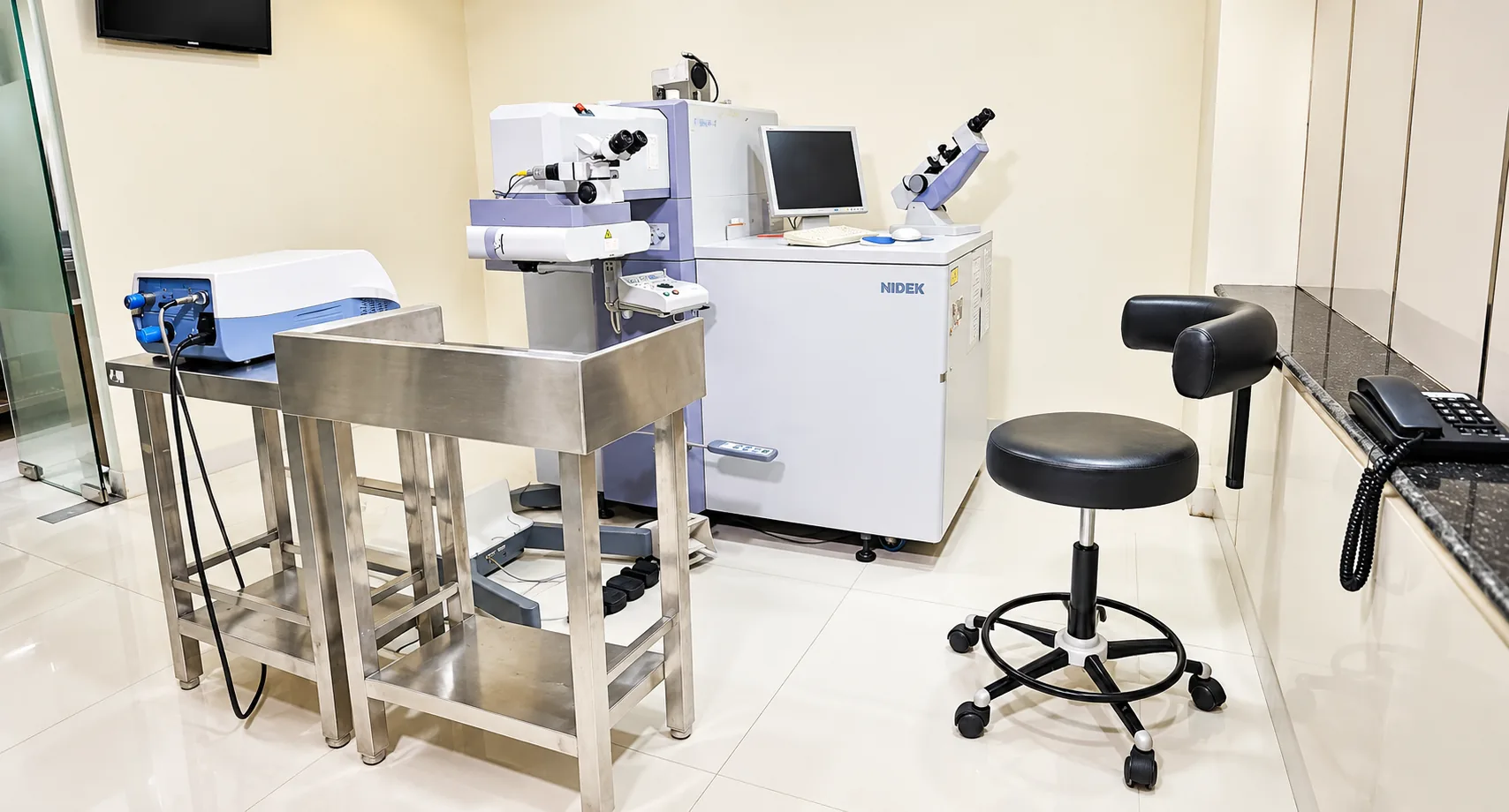
Ask whether a map shows a stable or changing pattern and how confident the team is in comparing two measurements. Contact lenses, dry eye and measurement quality can affect results, so preparation instructions matter. Request a copy of the report if you will seek another opinion or need serial monitoring. Technology can support decision-making but cannot replace a careful examination or rule out infection in a symptomatic eye. Confirm local equipment and appointment details directly with the hospital instead of assuming that every listed test is performed at every visit.
Preparing for an eye-surface appointment
Describe the main symptom, when it began and whether it is worsening. Mention contact-lens use, the type and wear schedule, cleaning solution, sleep or swimming in lenses, allergies, eye rubbing, previous infections or injuries, and all drops tried. Bring lenses and packaging if the clinician asks, but do not put a painful eye lens back in. If vision has changed, bring old spectacles and prescriptions so the team can compare them.
Ask whether the eye surface is intact, whether infection is possible, whether you need to stop contact lenses, which medicine is being recommended, and exactly when you should return. If a procedure is proposed, ask what it is expected to achieve and what it cannot do. Make sure you know which symptoms require an urgent call. If you have a sudden painful red eye or chemical exposure, seek urgent care instead of waiting for a routine appointment.
Protecting the cornea and knowing when to return
Follow the care plan exactly and avoid rubbing the eye. Use contact lenses only as directed; replace cases and solutions as recommended and never sleep or swim in lenses unless specifically advised by an eye-care professional. Do not share lenses, eye drops, towels or makeup. If a clinician has asked you to stop lens wear, wait until they confirm it is safe to restart. A surface can look or feel better before it is fully healed, so attend the planned check.
Get urgent help if pain intensifies, vision falls, redness spreads, light becomes hard to tolerate, discharge develops or a prescribed treatment is not helping as expected. After an injury, follow the specific directions supplied at the examination. Sudden changes should not be managed by increasing drop doses without medical advice. For long-term conditions such as keratoconus, keep comparison visits and bring prior maps; ongoing monitoring may detect a change even when day-to-day vision feels similar.
Safer contact-lens and eye-surface habits
Good lens hygiene lowers preventable irritation and infection risk. Wash and dry hands before handling lenses, use the disinfecting solution recommended for the lens, follow its replacement schedule, and keep the storage case clean and replaced as advised. Do not top up old solution, rinse lenses or cases with tap water, share lenses, or use saliva as lubricant. Avoid sleeping, showering or swimming in contact lenses unless the prescribing professional has specifically said it is appropriate. Ask the person who prescribed the lenses if care instructions are unclear.
Remove lenses and seek professional advice if the eye becomes painful, red, unusually light-sensitive or blurred. A contact-lens-related infection can worsen quickly, and an old antibiotic or steroid drop is not a safe substitute for examination. Keep lenses and the case available if the clinician asks to see them, but do not reinsert them into an irritated eye. Tell the clinician about the cleaning routine and when the lens was last worn; this can help assess what may have happened.
Protect eyes during activities where dust, flying particles, chemicals or tools could cause injury. Use suitable protective eyewear for the hazard. If something enters the eye, do not rub or use tweezers to remove an embedded object. For a chemical splash, start rinsing with clean lukewarm water immediately and seek emergency care. Injury can damage the cornea even when the eye looks only mildly red, so worsening pain or vision after an accident deserves prompt assessment.
Itching from allergy can encourage rubbing, but repeated rubbing is not a treatment and may aggravate discomfort. Ask a clinician or pharmacist what safe measures fit your symptoms, especially if you wear contact lenses or have a corneal condition. Do not share eye drops or use products marketed only to hide redness. A comfortable eye is not always a healthy eye; arrange an examination when symptoms persist, recur or affect vision.
Planning care for a changing corneal condition
Corneal conditions can change how glasses and contact lenses work. If a prescription changes repeatedly or vision remains distorted despite updated spectacles, ask whether the corneal shape should be examined. A lens that once felt comfortable may become difficult to tolerate if the surface or shape changes. Do not force a lens that causes pain. Tell the clinician about work, screen use, driving and other activities so that a proposed correction can be judged against real visual needs.
When keratoconus is suspected, ask whether the measurements show a stable shape or evidence of progression, how they compare with earlier results, and when they should be repeated. Ask what cross-linking is intended to do and whether it is suitable for the condition and stage in your own eye. It is not a shortcut to perfect uncorrected vision. A transplant is a major option reserved for selected situations; ask what less invasive approaches remain and what limitations or ongoing lens needs may continue afterwards.
For a surface infection or ulcer, understand which medicine is being used, when improvement should be apparent and what to do if it is not helping. Follow instructions about stopping contact lenses and returning for review. Some infections require close follow-up, so do not discontinue drops because pain settles before the prescribed course ends. If a prescribed medicine causes a reaction or you cannot use it as directed, call the clinician for advice rather than substituting a different bottle.
Protective habits matter especially after an earlier injury or in a person who rubs itchy eyes. Keep safety glasses available for cutting, grinding, gardening or handling cleaning products, and supervise children around sharp objects and chemicals. At work, use protection appropriate to the material and task. If an object is stuck in the eye, shield it without pressing and seek emergency help. If chemicals splash, rinse immediately and continue seeking professional care; do not wait for pain to become severe.
Knowing when a corneal problem is stable
Ask the clinician how they will know that a corneal problem has healed or stopped changing. A reduction in pain does not always mean an infection has cleared, and a stable-looking day does not establish that keratoconus measurements are unchanged. The care team may use symptoms, surface examination, repeat shape maps or other findings. Confirm which measurement is being compared and when the next check should happen. Bring the same records to a second opinion so that change over time can be assessed.
If a corneal condition affects both eyes differently, ask how to manage daily tasks safely while one eye is blurred or sensitive. Use the prescribed correction only as directed and ask what protective eyewear is appropriate at work or in sport. For any drop, confirm the bottle name and schedule; some patients use different medicines in each eye. Keep them in their original packaging and ask the pharmacist or clinician before using a bottle that looks different from the one prescribed.
Bring any previous eye reports, your current spectacles and a list of medicines or eye drops you use. Your clinician can explain which examinations or options are relevant to you.
Read the patient visit guideThis information is for general education and does not replace a personal consultation. Treatment recommendations vary from patient to patient and are made only after a detailed eye examination.