












The retina, macula and uvea
The retina is the light-sensitive tissue lining the back of the eye. It converts light into signals that travel to the brain. The macula is its central area and supports detailed central vision used for reading and recognizing faces. The uvea is a group of structures that includes the iris, ciliary body and choroid. Problems in these tissues can affect vision in different ways, so “retina and uvea” describes a broad area rather than one disease or one treatment.
Retinal conditions may be related to diabetes, blood-vessel changes, age-related macular disease, a retinal tear or detachment, inflammation, injury or other causes. Uveitis means inflammation inside the eye; it can have an infectious or non-infectious cause and may be associated with other health conditions. The same symptom can have several explanations. An examination is necessary to identify the problem, understand whether it is urgent, and decide if additional testing or referral is needed.
Symptoms that should never be ignored
Blurred or distorted central vision, difficulty reading, a dark or empty patch, changes in colour perception, or straight lines appearing wavy may indicate a macular problem. New flashes of light, a sudden shower of floaters, or a dark curtain or shadow at the edge of vision can be signs of a retinal tear or detachment. These symptoms need urgent same-day eye assessment; do not wait for a scheduled routine appointment or assume they are caused by age or fatigue.
Uveitis may cause pain, redness, light sensitivity, floaters or blurred vision, but symptoms depend on which part is inflamed and can sometimes be subtle. Diabetic retinal disease may cause no early warning at all. Record when a change began, whether it affects one or both eyes, and how quickly it is progressing. Sudden visual loss, severe pain, a curtain, or flashes with new floaters calls for urgent professional help. A home vision grid can help someone notice change but cannot diagnose or rule out disease.
Conditions that may affect retinal or uveal health
Diabetic retinopathy affects retinal blood vessels and may develop before a person notices symptoms. Diabetes can also lead to diabetic macular oedema, in which fluid affects central retinal tissue. Retinal vein problems, age-related macular degeneration, retinal tears and detachments have different causes and courses. Uveitis is inflammation inside the eye; some cases relate to infection, immune conditions, injury or no identified cause. The word “uveitis” does not by itself identify why inflammation has occurred or which treatment is appropriate.
Health history matters. Tell the ophthalmologist about diabetes and how it is managed, recent glucose or blood-pressure concerns if known, autoimmune or infectious illnesses, previous inflammation, injuries, surgery, medicines, and similar problems in the other eye. Eye findings may need to be considered alongside general medical care, and coordination with another clinician may be suggested. Do not stop a systemic medicine or use leftover steroid drops without medical advice; both the diagnosis and treatment can change with the cause.
How the retina and uvea are evaluated
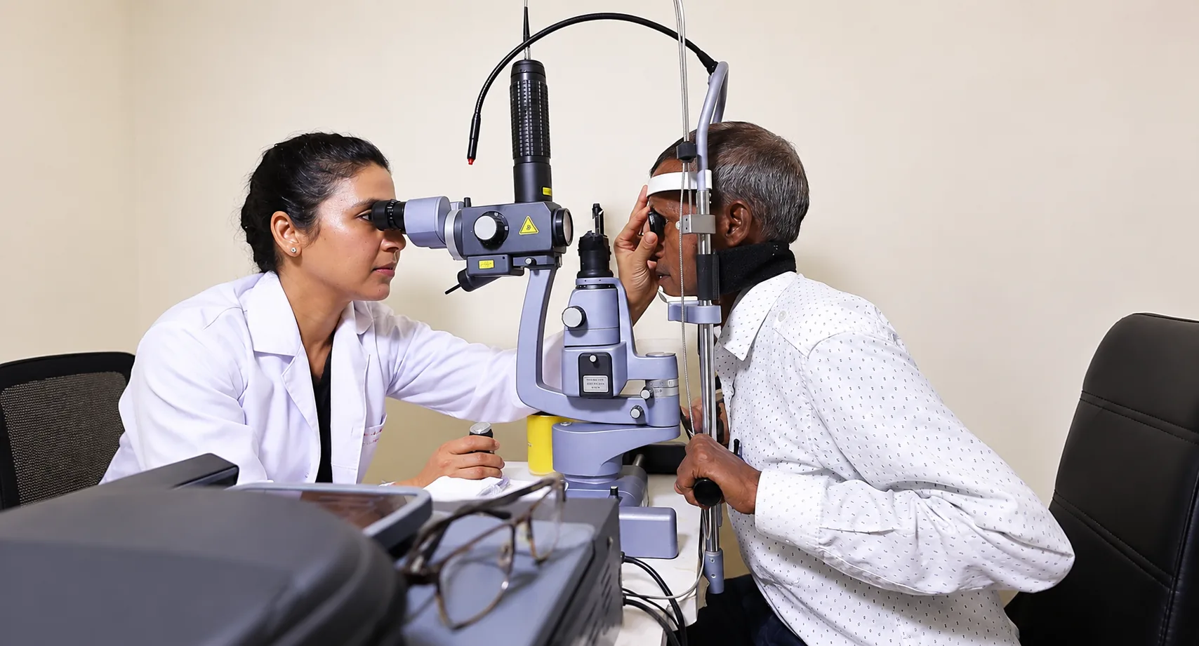
A clinician may check vision, examine the front of the eye, measure eye pressure and use dilating drops to inspect the retina. Depending on what is suspected, photographs, optical coherence tomography (OCT), retinal angiography, ultrasound or laboratory tests may be considered. No patient needs every test. Each examination answers a particular question; the clinician should explain what a test is expected to show and how the result could affect the plan. Dilation can temporarily blur near vision and increase light sensitivity, so ask what to expect before leaving.
For possible uveitis, the clinician may ask about general health and symptoms beyond the eyes. Blood tests or other investigations are selected according to the examination and suspected cause, not ordered automatically for everyone. Bring previous retinal images, eye reports, diabetes records if available, and a list of medicines and drops. Ask whether the condition is in the retina, macula or uvea, whether both eyes are involved, what urgent signs to watch for, and when results and follow-up should be discussed.
Treatment depends on the cause and stage
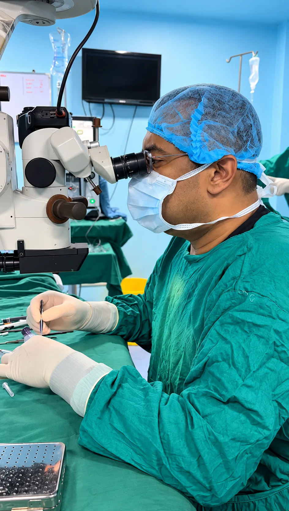
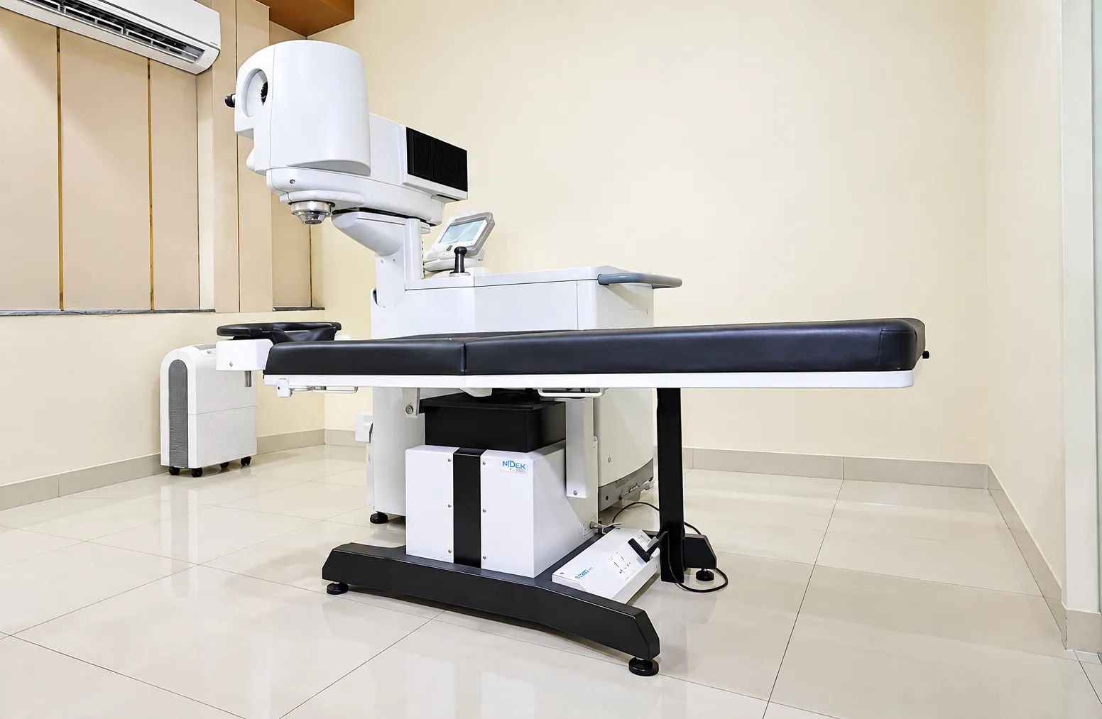
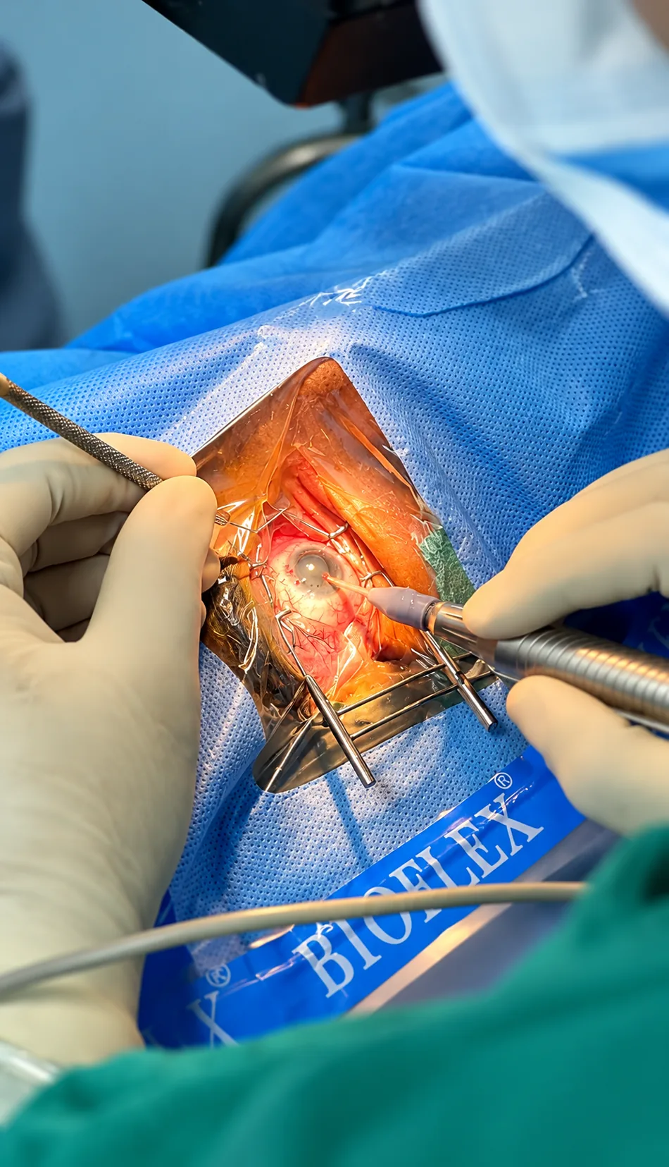
There is no single treatment for all retinal or uveal conditions. Depending on the diagnosis, care may include monitoring, treatment of an underlying health problem, injections into the eye, laser, or surgery. Uveitis treatment aims to control inflammation and address its cause; steroid medicines are one option but can have important risks and need professional supervision. A medicine that helps one inflammatory condition may be unsuitable for another, particularly when infection is possible.
For diabetic retinopathy, keeping regular diabetes care and retinal examinations matters even if vision feels normal. If a retinal detachment is suspected, treatment is urgent and is chosen by the retinal specialist according to the tear or detachment. Ask about the purpose of each proposed treatment, alternatives, expected number and timing of visits, side effects, what outcome is realistic, and what happens if treatment is delayed. Confirm the exact procedure and service offered at the hospital. No website can choose an injection, laser or operation for an individual eye.
Imaging helps answer focused clinical questions
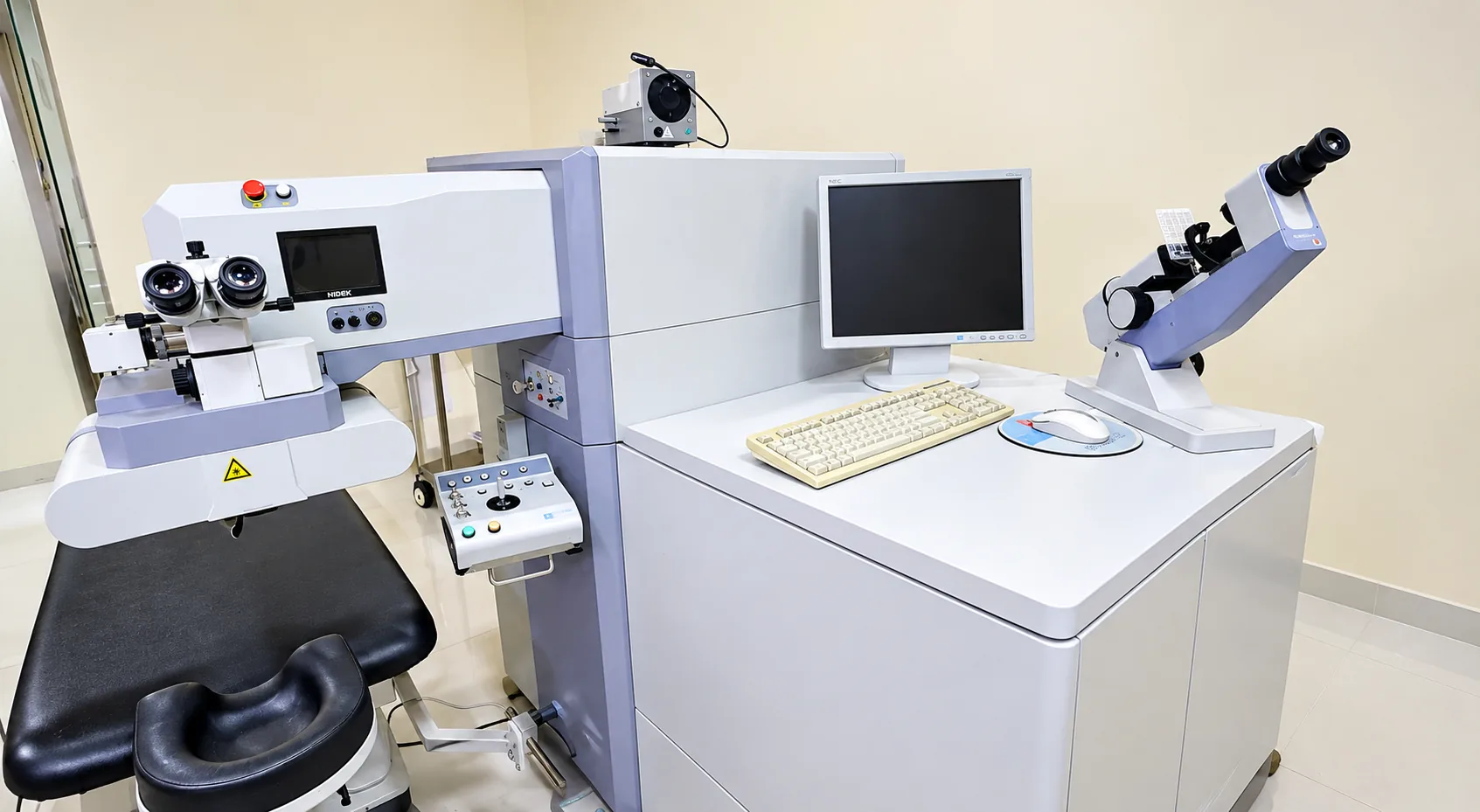
Retinal photographs can document appearance. OCT creates cross-sectional images that can help a clinician assess retinal layers and fluid. Other imaging or ultrasound may be helpful when a view of the retina is obscured or a particular vascular question needs answering. These tools provide information; they do not replace examination, symptoms and medical history. A normal result on one test cannot rule out every eye problem, and an abnormal image must be interpreted by a qualified clinician.
If the team recommends repeated imaging, ask what change they are monitoring and whether the result alters the treatment plan. Request an explanation of key findings in plain language and a copy of the report if available. Ask whether drops will be used, whether you can drive afterwards, and how results will be shared with the clinician managing diabetes or another systemic condition. Equipment availability varies by clinic; contact the hospital to verify whether a particular test is offered before arranging a visit.
Making a plan with your eye-care team
At the appointment, describe symptoms in order: what changed, when it began, whether it is getting worse, and which eye is affected. Mention diabetes, blood pressure, immune or infectious conditions, recent injuries, all medicines and past eye procedures. Bring reports and a support person if you need help remembering information. Ask the clinician to name the diagnosis or the possibilities being checked, whether the situation is urgent, and who will contact you with test results.
Before leaving, repeat the plan: treatment or monitoring, date of review, drops and precautions, symptoms that need urgent attention, and a number to call. If an injection, laser or surgery is proposed, request a separate conversation about benefits, uncertainty, risks, alternatives and consent. It is reasonable to ask for clarification or another clinical opinion when a recommendation is not understood. Do not postpone emergency assessment while seeking online reassurance about a curtain, sudden floaters and flashes, or sudden loss of vision.
Long-term monitoring and urgent care
Some retinal and uveitic conditions need repeated reviews even when symptoms improve. Keep the follow-up schedule and use drops only as prescribed. Do not miss a visit because a previous scan was stable: the care team may be looking for a change that is not yet noticeable to you. For diabetes, ask how often a dilated retinal examination is recommended for your situation and coordinate the plan with your diabetes-care team. Better systemic control can help protect health but does not replace eye screening.
Seek urgent assessment for a sudden increase in floaters, flashes, a shadow or curtain, abrupt distortion or central vision loss, or severe painful red eye with light sensitivity. If you receive an injection or procedure, follow the specific aftercare sheet and contact the treating team if pain, redness or sight worsens. Keep emergency contact details accessible. These are general education points: the clinician who has examined you should give individual instructions based on the condition, treatment and other health needs.
Keeping care coordinated between visits
Retinal and inflammatory eye conditions can involve several appointments, scans and health professionals. Keep a simple folder or phone note with the diagnosis, the eye affected, medicines and dose, procedure dates, imaging reports, next appointment and emergency number. Bring an updated list each time; eye drops count as medicines. If another clinician manages diabetes, blood pressure, infection or an immune condition, tell the eye team who they are and ask whether information should be shared.
For diabetes, retinal screening remains important even when vision feels normal. Ask the treating clinician how often the eye should be checked and how that schedule fits with the overall diabetes plan. Tell the team if appointments, transport or repeated visits are difficult; knowing the barrier may help discuss realistic arrangements. Do not miss or delay an injection because it is inconvenient without first speaking to the treating team. The interval is based on the disease and response, and changes should be medically advised.
If medicine is prescribed for uveitis or another inflammatory condition, understand its purpose, how to use it, how long it is planned for and what side effects to report. Do not restart a leftover steroid when redness returns: the cause may differ, and some infections can worsen with steroids. If you receive care from more than one doctor, show each clinician the complete medicine list and ask who is coordinating the overall plan. A clear contact person can reduce confusion when symptoms change.
Some treatments involve repeat visits rather than a one-time fix. Ask what improvement or stability means for your condition and what should happen if a visit must be rescheduled. Request an accessible written plan, especially if a caregiver helps with drops or transport. New floaters, flashes, a curtain or abrupt central vision change are urgent even if a specialist appointment is already booked. The new symptom may need assessment sooner than the planned review.
Preparing for repeat treatments and shared care
If an injection, laser procedure or operation is recommended, ask the clinician to explain the diagnosis first. Find out whether the goal is to reduce active disease, lower the chance of further damage, improve vision, or obtain information. These goals are not identical. Ask how response will be measured, how many visits might initially be needed, and what choices exist if the eye does not respond as expected. A treatment plan can change as scans and symptoms change; this does not mean that one appointment predicts the entire course.
Discuss discomfort, infection risk, medication interactions, transport and help at home. Confirm whether you should continue routine medicines and drops; never stop a medicine used for diabetes, blood pressure or immune health without the clinician who prescribed it. If you need an interpreter or an accessible written schedule, ask before the appointment. Keep procedure instructions with your medicine list and the phone number for reporting a problem. After treatment, follow the instructions for that specific procedure, not advice for someone else’s injection or laser.
Uveitis may prompt questions about general health because eye inflammation can sometimes be related to infection or a condition elsewhere in the body. This does not mean that every person needs broad testing. Your clinician selects investigations based on the pattern, history and examination. Tell the eye team about fever, skin changes, joint symptoms, recent infections and previous diagnoses when relevant; share results from other doctors if available. Ask who will explain test results and whether you should continue care with a general physician or another specialist.
Ask for a clear division of responsibilities when more than one team is involved: who prescribes each medicine, who monitors side effects, who orders imaging and whom to call if vision changes. This is especially helpful when a person sees different services for diabetes and retinal care. If the plan feels confusing, ask for one concise written summary. Clear coordination helps patients attend the right review at the right time and reduces the risk of stopping treatment or repeating a medicine by mistake.
Leaving each visit with a clear action plan
Before leaving, ask the care team to state the next action and its timing: another examination, an image, a medicine change, an injection, a procedure or observation. Ask who will review any pending test and how the result will be communicated. If an appointment is made for a specific treatment window, write down the date and number to call if a delay becomes unavoidable. Do not use a general online schedule to decide whether to postpone an appointment chosen for your own retinal findings.
If several family members help, choose one person to keep the current medicine list and appointment calendar, with the patient’s permission. Bring the list to every eye and medical visit, and remove medicines that have been stopped only after confirming with the prescribing clinician. If the reason for a change is unclear, ask which diagnosis or test prompted it. This small routine reduces confusion between similar eye drops and supports safe follow-through across teams.
Bring any previous eye reports, your current spectacles and a list of medicines or eye drops you use. Your clinician can explain which examinations or options are relevant to you.
Read the patient visit guideThis information is for general education and does not replace a personal consultation. Treatment recommendations vary from patient to patient and are made only after a detailed eye examination.