










The macula and detailed central vision
The macula is the central part of the retina, the light-sensitive tissue at the back of the eye. It supports detailed vision used for reading, recognising faces, seeing fine print and identifying central detail. Macular conditions affect this central area, but they do not all have the same cause or course. They can arise with age-related macular degeneration, diabetes, inherited conditions, inflammation, circulation changes or other retinal problems. A patient needs an examination to know which explanation is relevant.
The aim of a macular assessment is to understand what is causing the visual change, how the central retina is affected, and whether monitoring or treatment is appropriate. Some macular changes are slow; others need prompt attention. A diagnosis should be explained in plain language, including what is known, what remains uncertain, and what the next review is intended to detect. Information about one person’s injection, supplement or scan schedule should not be copied as advice for someone else.
Recognising central vision changes
People may notice that straight lines appear bent or wavy, words look distorted, a patch in the centre seems blurred or missing, colours look different, or reading takes more effort. Faces may be harder to recognise even though side vision seems intact. Changes can affect one eye before the other, so the unaffected eye may compensate and hide a problem. Cover one eye at a time only as a simple way to notice a difference—not as a test that can identify its cause.
New distortion or loss of central vision should be reported promptly, particularly if it begins suddenly or changes quickly. Do not wait for pain: many retinal conditions are painless. Sudden floaters, flashes or a curtain-like shadow point to a different urgent retinal concern and need immediate assessment. A grid or online image may help someone describe what they notice, but it cannot diagnose macular disease or establish that the retina is stable. The clinician’s examination and imaging are needed.
Different causes need different plans
Age-related macular degeneration (AMD) is one well-known macular condition and is commonly discussed as dry or wet (neovascular) AMD. The two forms differ, and the presence of AMD does not mean that a person will become blind. Diabetes can cause fluid to collect in the macula; inflammation, retinal vascular problems, inherited disorders and other disease can also affect central detail. Age, smoking history, family history, diabetes and prior retinal conditions may be part of the conversation, but none alone confirms a diagnosis.
Treatment varies according to the specific condition, its stage, the eye involved, prior response and the person’s overall health. Some findings are monitored; others may be treated or referred. Vitamins or supplements studied for certain stages of AMD are not a general prevention pill and can be unsuitable for some people. Ask whether evidence applies to your stage and whether any supplement could interact with your medicines. Do not start a high-dose product based on an advertisement or someone else’s experience.
How clinicians assess the macula
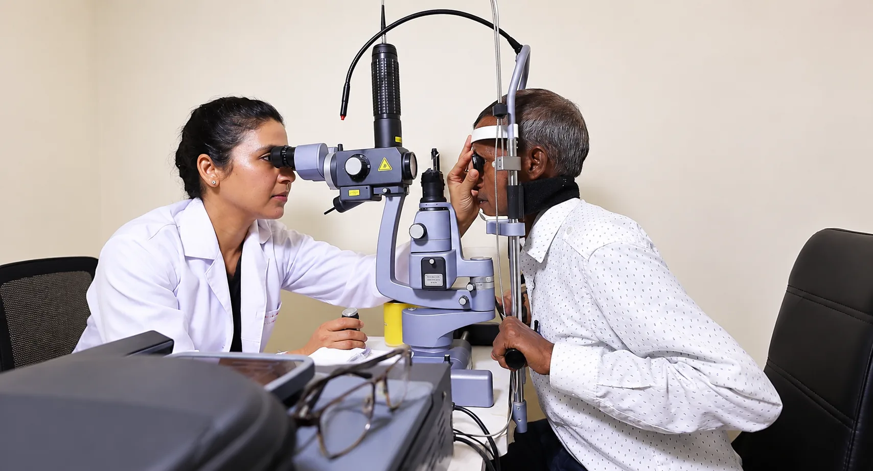
The assessment usually includes a vision check and a dilated examination of the retina. Imaging may include retinal photographs and optical coherence tomography (OCT), which creates a cross-sectional view of retinal layers. Some conditions need other imaging. A clinician uses these findings alongside symptoms, previous scans and health history. One scan is a record at a point in time; comparison with prior images may be more informative than an isolated result.
Bring older OCT or retinal images if available, previous letters, a current medicine list and details of relevant conditions such as diabetes. Mention which eye seems affected and whether straight lines look different. Ask the clinician to identify the macular change in plain language, whether the other eye should also be monitored, and how soon results need review. If dilation is used, near vision and light sensitivity can temporarily change; check whether you should bring sunglasses or avoid driving until vision returns.
Treatment and support depend on the diagnosis
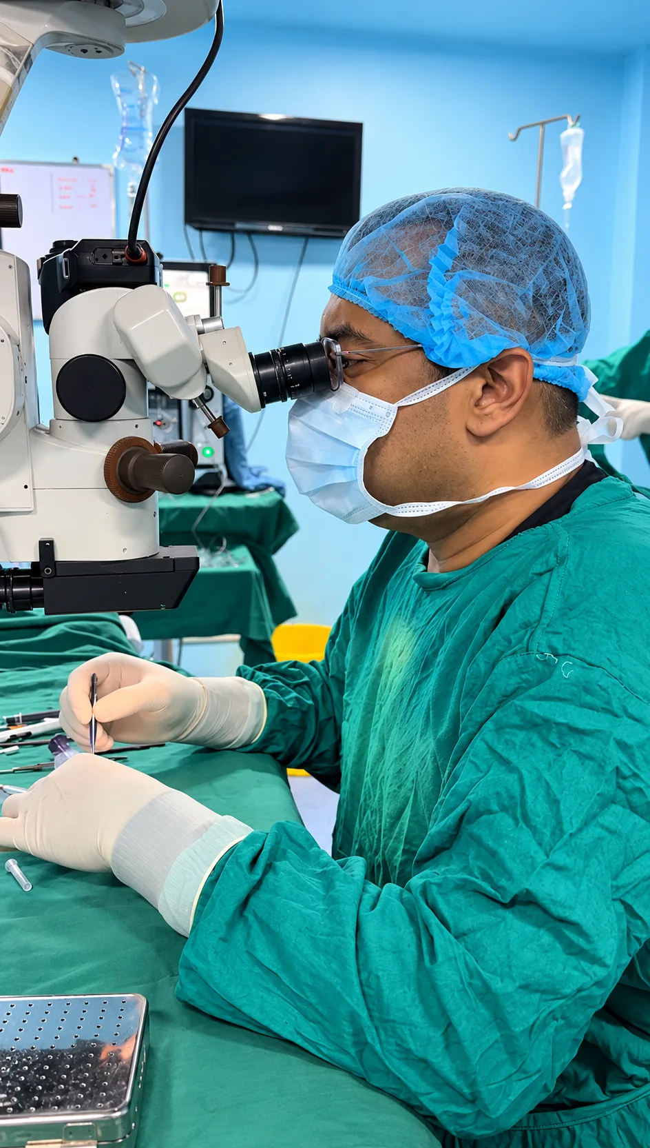
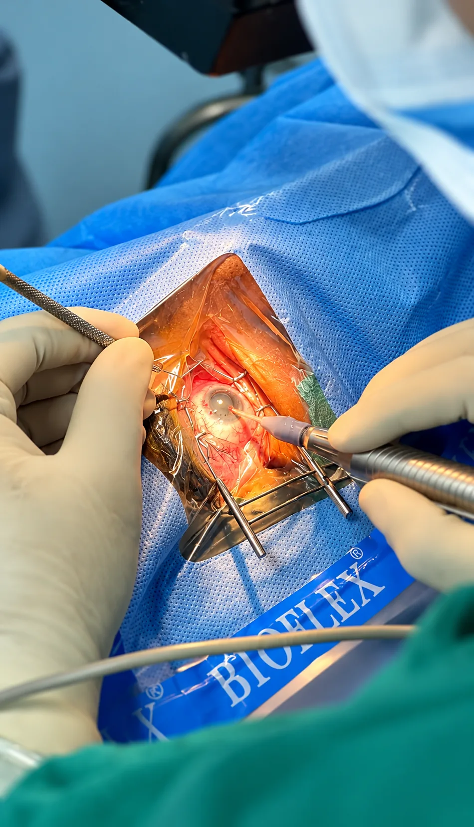
Not every macular finding requires an injection or operation. Depending on diagnosis and activity, a clinician may recommend observation with scheduled imaging, treatment of a contributing condition, injections inside the eye, laser in selected situations, or another specialist service. For wet AMD and some other active macular diseases, anti-VEGF medicines injected into the eye may be considered. The number and interval of injections are individual; ask why this medicine is proposed and how the response will be assessed.
Discuss benefits, risks, alternatives, costs, likely visit schedule and the consequences of delaying care. Ask how the procedure is performed, what to expect afterwards, which symptoms call for immediate contact, and how the other eye will be monitored. If central vision remains limited after treatment, low-vision support, magnification, contrast adjustments and lighting changes may make tasks easier. Ask for practical recommendations tailored to your vision rather than assuming that treatment will fully restore sight.
Using scans to track change over time
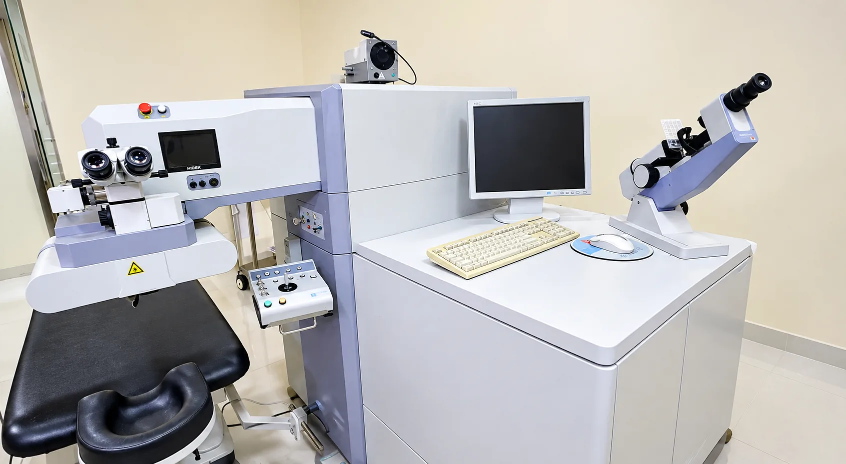
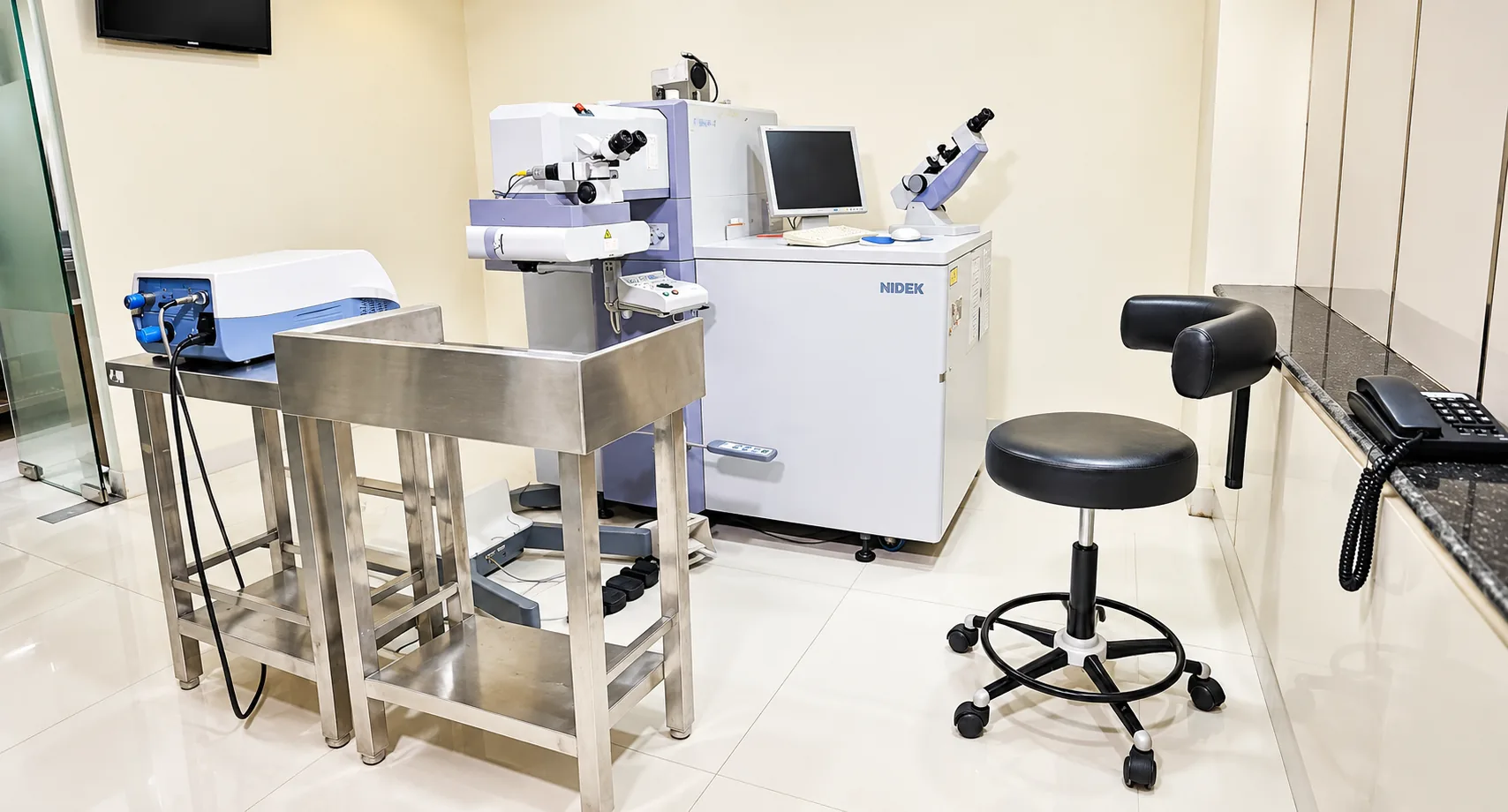
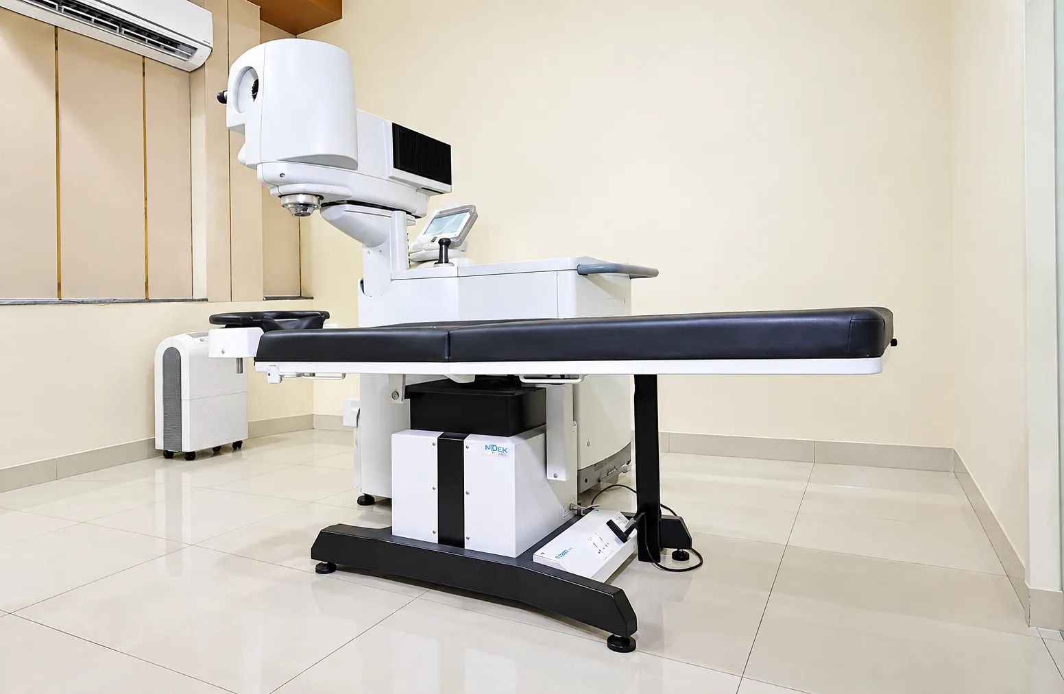
OCT and fundus photographs can help document features of the macula. An OCT image may show retinal thickness, fluid or layers that are difficult to describe during a short appointment. Imaging helps a clinician compare change and judge response to treatment, but the image is not a diagnosis on its own. Scan quality, eye movement, eye anatomy and the question being asked all matter. Ask the clinician to show or describe the finding and explain how it relates to your sight.
Different visits may use different tests. There is no need to request every test seen online; a test is useful when it helps answer a clinical question. Ask how soon results will be reviewed, whether a new symptom should bring the visit forward, and whether a scan has to be repeated at the next appointment. If you receive copies, keep them dated with the corresponding clinical note. Confirm the hospital’s specific imaging availability with the care team before an appointment.
Preparing to discuss diagnosis and goals
Before an appointment, note what tasks are difficult and what changes you have noticed. Bring glasses, prior retinal reports and images, medicines and relevant health records. If someone helps with reading or transport, consider inviting them to listen, while keeping the patient’s own priorities central. Explain whether the main goal is to read, drive, work, recognise faces, or manage another everyday task. That helps the clinician describe what a proposed treatment may realistically achieve.
Ask: What is the exact diagnosis? Is it active or stable? What is the plan and when should it be reviewed? Which symptoms should prompt an earlier call? Will treatment preserve vision, improve it, or mainly reduce the chance of further change? Answers may be different for each eye. If you are offered an injection or another treatment, ask about the full consent process and aftercare. You are entitled to understand why a plan is recommended and what other reasonable options exist.
Ongoing monitoring and practical vision support
Keep the review schedule even when the eye feels comfortable. Macular disease can change without pain, and treatment is often more useful when a clinician can compare symptoms and scans over time. Use any prescribed eye drops exactly as directed, but do not add supplements or medicines without checking. For diabetes-related macular disease, keep both retinal appointments and general diabetes care. A stable reading on one visit does not mean future checks are unnecessary.
Tell the eye team promptly if distortion, a dark patch or central blur newly appears or worsens. Sudden floaters, flashes or a curtain need urgent assessment for possible retinal tear or detachment. If vision remains reduced, ask about low-vision rehabilitation and everyday adjustments: larger print, task lighting, high-contrast labels and screen magnification can help some people. These supports do not replace medical assessment; they are ways to maintain independence while a clinician manages the eye condition.
Adapting everyday tasks when central vision changes
Changes in central vision can make familiar tasks unexpectedly tiring. Add light to the page or work surface while reducing reflections, enlarge print on a phone or computer, and increase contrast between labels and backgrounds. A magnifier or screen reader may help with specific tasks. Move loose rugs and clutter from walking routes, keep frequently used items in a consistent place, and use large, clear labels on medicines. These adjustments can improve access but do not diagnose the reason for a change or replace planned retinal care.
Tell the clinician which tasks matter most rather than asking only whether the scan looks better. Reading a bill, cooking, recognising a face, using public transport or returning to work may each require different support. Ask whether a low-vision service, occupational therapist or rehabilitation professional can assess your needs. Do not assume support is useful only when sight is extremely poor; practical advice can be valuable while a person is still receiving medical care.
If you use an Amsler grid or another home monitoring tool, follow the clinician's instructions about how to use it and what change should prompt a call. A grid is not an eye test, and a normal-looking result cannot rule out disease. If lines suddenly become more distorted, a new blank patch appears or central vision falls, contact the eye team promptly. Do not wait for the next scan simply because the home chart seems unchanged.
Bring a family member or trusted person to a visit if you want help remembering information. Ask the clinician to describe each eye separately and write down the diagnosis, plan, medicine changes and review date. If the explanation uses unfamiliar terms, ask what findings mean for your activities. You have the right to make decisions after understanding benefits, uncertainties, alternatives and follow-up. Support with daily tasks and medical care can proceed together; one does not replace the other.
Understanding monitoring, treatment and uncertainty
The word “macular” describes a location, not a single diagnosis. Ask the clinician to name the condition, whether it is active, which eye is affected, and whether a treatment is intended to improve current vision or reduce the chance of further change. A stable scan may support observation in one situation and still require repeat checks in another. If treatment is recommended, ask how the team will judge whether it is working and what finding would lead to a different approach.
If injections are discussed, ask what medicine is proposed, how it is given, the likely visit pattern, the main risks and the symptoms that need a same-day call afterwards. Ask whether observation or another option is reasonable and what could happen if treatment is delayed. Do not assume that another patient’s injection schedule applies to you; age, diagnosis, scan findings and response differ. Keep every appointment or call the clinic early if transport, cost or caregiving makes attendance difficult, so the care team can advise safely.
Some people hear about vitamins or supplements after an AMD diagnosis. Research evidence applies to certain categories and stages; it does not mean every person should take the same product. Tell the ophthalmologist about all vitamins and medicines before starting a supplement, particularly if you smoke or have other health conditions. Ask whether the evidence applies to your findings, what amount is appropriate if one is recommended, and whether the product could interact with another treatment. Food and supplements should not be used to delay evaluation of new distortion.
Keep a dated record of visual changes and test results. If a new distortion appears between appointments, note when it began and contact the care team rather than waiting to document it at the next visit. Home grids can help describe change but are not a clearance test. Encourage family members to ask before taking over tasks; many people with reduced central vision remain capable of independent decisions and benefit most from practical tools that preserve control over daily life.
Turning scan results into understandable next steps
When the clinician reviews an image, ask what finding has changed since the last visit and whether the change matters for the treatment plan. A technical term can sound alarming without context; ask whether it describes location, severity or activity. Check whether the result is for the right eye, the left eye or both, and what symptoms you should monitor between visits. If you receive a printed scan, keep the date and the clinical explanation with it so that a future provider does not interpret an image without its history.
If the plan is observation, ask exactly when to return and what change should prompt an earlier call. If treatment is planned, confirm who schedules it, what to do with regular medicines, and what follow-up is expected. If practical barriers threaten attendance, say so promptly; a clinic may be able to discuss options, but only the treating team can advise how a delay affects your case. Understanding the next action is more useful than trying to interpret an image alone.
Bring any previous eye reports, your current spectacles and a list of medicines or eye drops you use. Your clinician can explain which examinations or options are relevant to you.
Read the patient visit guideThis information is for general education and does not replace a personal consultation. Treatment recommendations vary from patient to patient and are made only after a detailed eye examination.